X-Ray
How a dental cavity is visible in x-ray?
Dental cavity is technically called as dental caries, is a common complaint with which the patient lands up in a dental setup. Usually the cavity starts in the tooth as a black or brown spot in the tooth structure. At this stage, even the common public is not aware that the cavity is started in the tooth. The reason behind is; there is no pain, sensitivity, discomfort in the starting stage. When the cavity progress to a moderately deep level which we call as progression into the dentin. The cavity starts to appear visible in the x-ray. We technically tell as, when there is more than 30% of loss of mineral content in the tooth there is evidence on the X-ray as black darkening.
Inside every tooth there is a pulp chamber giving life to the tooth. This chamber highly innervated with veins & nerves. When the cavity touches the pulp, the individual faces severe pain. When the X-ray taken at this stage, we clearly visualize the cavity is clearly touching the pulp. This is the common scenario seen in the dental clinic, at this stage of problem ideally the dentist advices Root canal treatment (or) extraction. If the patient willing to save the tooth with Root canal treatment, a prosthetic crown is suggested after Root canal treatment. In children, the distance between the top layer of crown to the pulp chamber for back teeth is casually around 1.5 - 1.7 mm so the progression of decay in children is very quick, landing up with severe pain.
The protocol for dental treatment varies from adult. Kindly have a dental checkup done with your reliable dentist to have cavity free smile. Only the dentist will be able to detect the cavity at early stage of life.
Decay in-between teeth
Decay in-between teeth is also called proximal decay or caries, is again a common scenario in many children’s & adults who develop dental cavities. The reason behind this problem is, whenever we eat food there is a thin layer called Dental plague will accumulate in between teeth. If left unattended, it start slowly settling. This material bio-film is a good environment for the cavity causing bacteria to sit, stay & multiply. If the plague is not effectively cleaned, it can start developing into decay. When the damage process is initiated it not only affects one but both the tooth (i.e.) the back side of one tooth & front side of other tooth, which is called DO & MO in dentistry. When these lesion starts happening in the starting stage usually there is no discomfort, sensitivity or pain. As the decay progress, the first sign of the problem is food trapping in-between teeth, for which the individual starts using tooth pick to remove food. When the severity worsens, the tooth structure on the top breaks open because of the underlying weak structure and slowly discomfort starts setting in the patients teeth. At this gesture, patient usually visit the dentist for dental checkup to be done. When the client report with this problem, an x-ray is taken to rule out the severity of the problem if the decay is confirmed either a filling, inlay / onlay, RCT with full coverage crown is suggested based on the severity of the problem.
Gum problems in adults
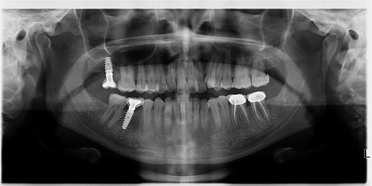
A very common situation in which the patient reports to us, with a bleeding gums. In this situation the dentist evaluates the teeth & gums. The gums are more specifically evaluated with Williams problem to measure the depth of pocket. If the pocket dept is more than 5mm, blood ensures if the problem is generalized then the patient is advised for an OPG. This evaluates, the general bone condition of teeth. OPG x-ray reveals general health of the bone surrounding the tooth called the ‘‘alveolar bone’’. If the gum problem is very bad, the height of the alveolar bones is decreased in a condition called ‘‘chronic periodontitis’’ were mobility is commonly seen in patients. If the gum condition is healthy, the bone level is also good capturing the tooth to the neck level. So OPG play a major role is assessing the surrounding bone condition of the tooth.